Your GI Center
Info
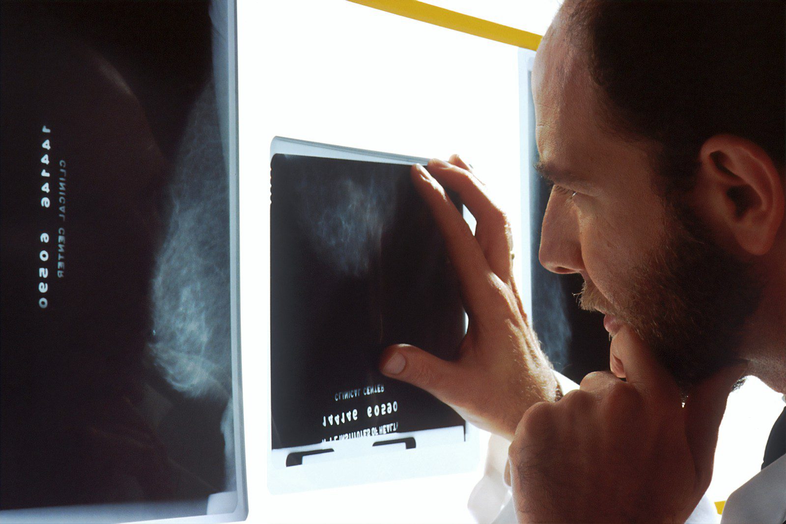
Endoscopic ultrasound is a minimally invasive procedure that uses a specialized endoscope equipped with an ultrasound transducer at its tip. This unique combination allows gastroenterologists to obtain detailed, high-resolution images of the digestive tract wall and surrounding structures, including the pancreas, bile ducts, liver, gallbladder, and nearby lymph nodes. The ultrasound component provides images from inside the body, offering superior visualization compared to external ultrasound or other imaging modalities.
The EUS procedure involves inserting the echoendoscope through the mouth (for upper EUS) or rectum (for lower EUS), positioning it close to the area of interest. Because the ultrasound transducer is placed directly adjacent to the target organs, EUS provides exceptionally detailed images with resolution superior to CT scans, MRI, or external ultrasound. This proximity allows for accurate assessment of tissue layers, detection of small lesions, and precise measurement of abnormalities.
EUS is considered the gold standard for evaluating pancreatic masses, cysts, and inflammatory conditions. The procedure can differentiate between solid masses and cystic lesions, assess the relationship between pancreatic abnormalities and surrounding structures, and help determine the likelihood of malignancy. EUS is particularly valuable for detecting small pancreatic tumors that may not be visible on other imaging studies and for evaluating chronic pancreatitis changes.
Accurate staging of gastrointestinal cancers is crucial for determining the most appropriate treatment approach. EUS provides detailed information about tumor depth (T-stage), involvement of nearby lymph nodes (N-stage), and relationship to surrounding structures. This information is essential for surgical planning, determining resectability, and selecting appropriate treatment strategies for esophageal, gastric, rectal, and pancreatic cancers.
EUS excels in evaluating submucosal lesions, which are abnormalities beneath the surface lining of the digestive tract that cannot be adequately assessed by standard endoscopy. The procedure can determine the exact layer of origin, measure the size and characteristics of these lesions, and help differentiate between benign and potentially malignant conditions. This information is crucial for determining whether intervention is necessary.
EUS provides excellent visualization of the bile ducts, gallbladder, and surrounding structures. The procedure can detect bile duct stones, assess biliary strictures, evaluate gallbladder abnormalities, and identify causes of biliary obstruction. EUS is particularly useful when other imaging studies are inconclusive or when detailed assessment of biliary anatomy is required.
One of the most significant advantages of EUS is its ability to obtain tissue samples from deep-seated lesions through fine needle aspiration. Under direct ultrasound guidance, a thin needle is passed through the endoscope and into the target lesion to collect cells or tissue for microscopic examination. This technique provides definitive diagnosis for pancreatic masses, lymph nodes, and other abnormalities while avoiding more invasive surgical procedures.
EUS-guided fine needle biopsy uses specialized needles to obtain larger tissue samples compared to standard FNA. This technique provides more tissue for histologic analysis and molecular testing, which is increasingly important for personalized cancer treatment decisions. FNB is particularly valuable for pancreatic lesions where tissue architecture and additional testing are crucial for accurate diagnosis.
Advanced EUS techniques include drainage of pancreatic pseudocysts, celiac plexus neurolysis for pancreatic cancer pain, and biliary drainage in selected cases. These therapeutic applications of EUS provide minimally invasive treatment options for complex conditions that previously required surgical intervention.
EUS preparation is similar to upper endoscopy, requiring fasting for at least eight hours before the procedure. Patients should not eat or drink anything after midnight before a morning procedure or for eight hours before an afternoon procedure. Blood tests may be required, particularly if tissue sampling is planned. Our medical team will review all medications and provide specific instructions about which medications to continue or discontinue before the procedure.
EUS is performed under conscious sedation to ensure patient comfort throughout the procedure. The examination typically takes thirty to ninety minutes, depending on the complexity of the case and whether tissue sampling is performed. The gastroenterologist carefully guides the echoendoscope to the area of interest and obtains detailed ultrasound images from multiple angles and orientations.
If fine needle aspiration or biopsy is indicated, the procedure is performed under direct ultrasound guidance to ensure accurate targeting and maximize diagnostic yield. Multiple passes may be necessary to obtain adequate tissue samples, and on-site cytologic evaluation may be available to confirm adequacy of the specimens obtained.
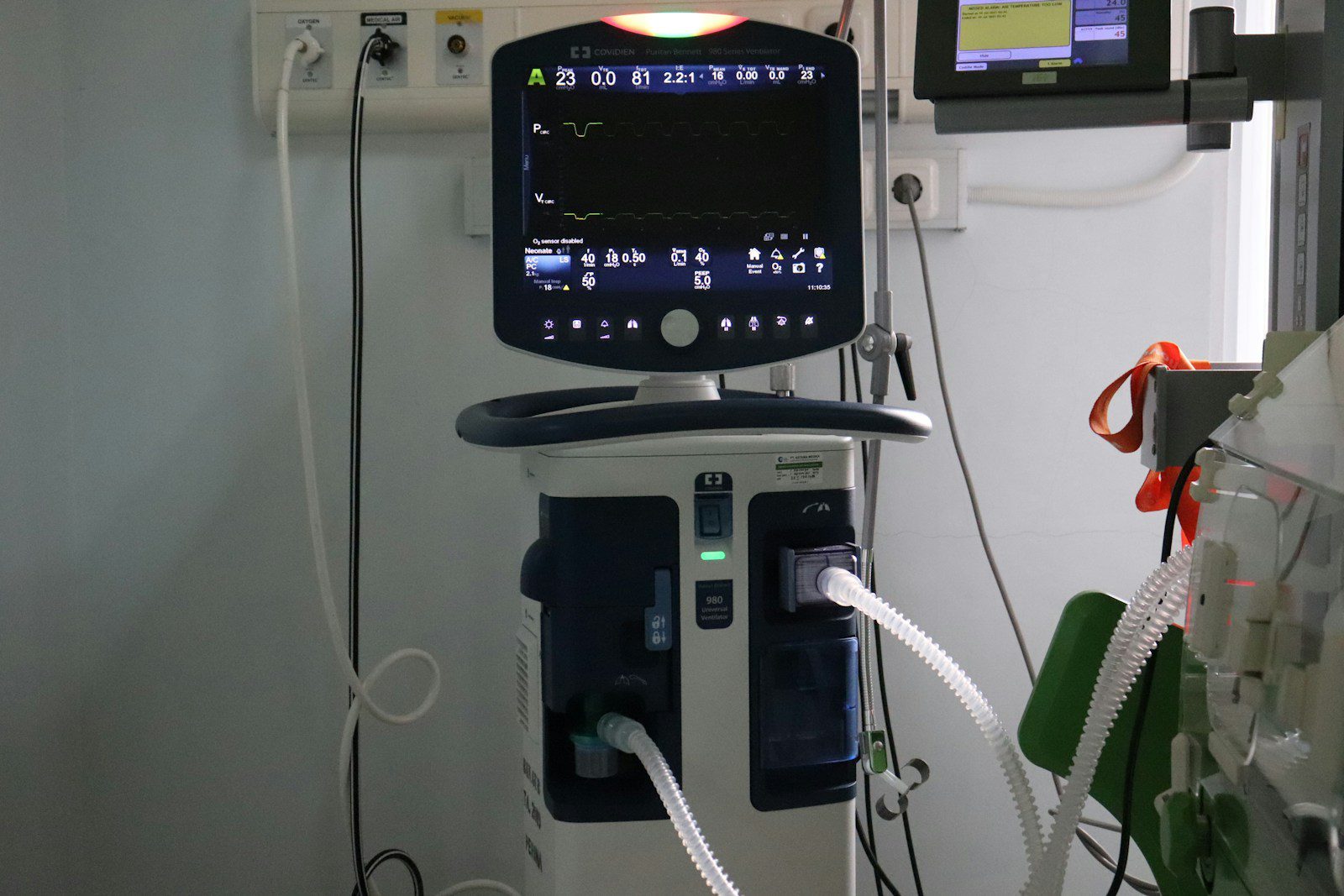
Your GI Center utilizes state-of-the-art EUS equipment with high-frequency ultrasound transducers that provide exceptional image quality and resolution. Our endoscopy centers in Houston and Lake Jackson are equipped with advanced EUS systems that offer both radial and linear imaging capabilities, allowing for comprehensive diagnostic evaluation and precise tissue sampling when indicated.
Your GI Center utilizes state-of-the-art EUS equipment with high-frequency ultrasound transducers that provide exceptional image quality and resolution. Our endoscopy centers in Houston and Lake Jackson are equipped with advanced EUS systems that offer both radial and linear imaging capabilities, allowing for comprehensive diagnostic evaluation and precise tissue sampling when indicated

Both Dr. Meah and Dr. Siddiqui have received extensive training in EUS techniques during their gastroenterology fellowships at prestigious medical institutions. Their combined experience in performing hundreds of EUS procedures ensures that patients receive the highest quality care and most accurate diagnostic information available.
12951 South Freeway Houston
TX 77047
(713) 436 8171
Advanced EUS capabilities with tissue sampling and therapeutic interventions.
720 Avenue F North Bay City
TX 77414
(979) 292-0033
EUS consultation and follow-up care for Matagorda County patients.
109 Parking Way
Lake Jackson, TX 77566
(979) 292-0033
Comprehensive EUS services for the Brazosport area.

After EUS, patients recover in our comfortable observation area while the sedation effects wear off. Most diagnostic EUS procedures allow patients to go home the same day, typically within one to two hours after the procedure. If tissue sampling was performed, patients may experience mild throat discomfort or abdominal soreness, which usually resolves quickly.
Results from the ultrasound examination are typically available immediately and will be discussed with patients before discharge. If tissue samples were obtained, results from pathologic analysis are usually available within a few days to one week, depending on the type of testing required. Our medical team provides comprehensive follow-up care and helps coordinate additional treatments or referrals as needed based on EUS findings.

We are available 24/7. Call Now for Immediate Assistance!
For advanced diagnostic imaging and tissue sampling of gastrointestinal and pancreatic conditions.
Call 1-888-292-0010 to discuss EUS evaluation with our specialists.